
SIMPLE AND SAFE METHOD FOR THE REMOVAL OF EAR WAX
 |
Fernando R. Kirchner MD. Ex tenured Professor Ear, Nose and Throat and Head and Neck Surgery Retired Clinical Associate Professor of Family practice Midwest University Medical School. USA Fellow of American College of Surgeons. Ex Member NIH Study Group Web site: https://www.ferdsjourney.com |
Our first aim for this paper is to review the structure of the human ear canal, describe its functions, and outline a safe and economical method of wax removal that is available to the public.
Anatomically the ear canal is formed by a deep bony tube that ends at the eardrum, and a peripheral cartilaginous portion that is part of the auricle.
The ear canal’s two functions are: first, to keep this passage open to permit air sound waves to reach the ear drum, and second, to clean itself by an automatic cleansing mechanism.
These two functions are very important to humans and other species, because without them, hearing would be significantly impaired, due to the fact that air sounds, and sounds in liquids, such as the fluids of the inner ear, have different acoustic properties. This impairment clearly is experienced when one tries to hear under the water, and it is overcome by the trasducer effect of the tympanic membrane and the small bone located in the middle ear.
Since the ear canal is a closed canal, its self cleansing mechanism is equally contributory to this function. This property is produced by the constant cellular migration from the surface of the ear drum to the outside walls of the ear canal. It suffices to paint a dot of India ink on the surface of the ear drum to demonstrate by repeated observations, the migration of these dots to the periphery of the canal. Additional information on weekly ads of pharmacy stores.

SIMPLE PRACTICAL METHOD FOR THE REMOVAL OF EAR WAX.
In contrast to the bony deeper portion of the ear canal that requires for its examination special illumination and instrumentation; the external cartilaginous segment of this canal can be manipulated, and is easy to inspect.
Anatomically speaking, this cartilaginous portion has hair follicles and special wax glands, suggesting that the added function of this segment is to protect the ear canal from the entry of external foreign bodies from the outside.
However, at times dry wax or dry wax together with hair and dry skin, can plug the ear passage, sometimes in a formidable fashion.
Initially the obstruction is only manifested by hearing loss. However, these plugs can grow deeper into the sensitive boney ear canal, or get wedged against the ear drum. Although diving may contribute to this wedging, incorrect attempts to remove these plugs are also capable of producing this complication.
A kit is available to the non-specialist and the public at large to correct these problems.
 |
Through some years of clinical research, NeilMed has developed the “Clearcanal, Dr. Mehta’s Ear Wax Removal Complete Kit.” that is available in most drug stores in the USA. I personally have seen patrons obtain this kit at our local CVS and Walgreen pharmacies. There are three components in the kit, a drop bottle containing a specially formulated ear wax softener, 2 quality ear plugs used to keep the softener inside the canal for ten minutes , and third, a pressurized spray can that is used to dislodge by flushing the already softened wax plug or plugs. The instructions in the package, of how to use the kit has been simplified by “ Dr. Mehta’s by the rule of ten: Rule of 10 ”Method for Ear Wax Removal,” 10 DROPS per ear, plug ears for 10 MINUTES, rinse each ear for 10 SECONDS. The kit contains 10 complete treatments. |
There is also a plastic cup that is used to collect the returning flush from the pressurized can.
After trying this kit a number of times, I would like to add 3 personal observations.
1. – Use the pressurized can to flush out the wax plugs and debris, WITHOUT OBLITERATING THE RETURN FLOW. This will avoid creating unwanted pressures in the ear canal that might damage the ear drum.
2. – One may aid the flushing to help to dislodge the wax, by moving the auricle in a circular fashion.
3. – If the collecting cup gets in the way of the flush, use instead a towel draped around the neck.
All the above steps should be carried out in a well illuminated area.
Personally, I prefer the sitting up position, looking at the ear in a straight line, holding the flushing can in one hand, and the auricle with the other hand.
After the flushing, the ear is dried with a towel. Tilting the head, and mobilizing the auricle, is usually sufficient for drying off the rinse solution.
Finally, at times patients with impaction in ear canals that are extremely convoluted and narrow need to consult a specialist for the removal of these impactions.
Find similar sort of waxes on the CVS Pharmacy and CVS Weekly Ads. Also read the reviews to learn cheapest options.
PIONEER STUDIES ABOUT THE IMPAIRED CLEANSING MECHANISM OF THE EAR
Papanicolaou OR Pap Smears of the ear canal
Deeper to the cartilaginous portion of the ear canal is the bony portion. This segment is closed by the Tympani membrane. The entire ear canal is covered by squamous epithelium, including the Tympanic membrane, where the epithelial layer is very thin.
This layer is constantly renewed. This replenishment produces cellular debris that needs to be evacuated.
Miraculously, the canal has a built in cleansing mechanism. This mechanism consists of an automatic cellular stream directed to the outside, thus avoiding the accumulation and impaction produced by these discarded elements.
The study of this cleansing mechanism provided the author with information on how to timely resolve issues that otherwise would lead to the destruction of the ear.
Cases in point are pockets of retraction or invagination that occurs on some tympanic membranes. In the majority of these invaginations the cleansing mechanism is not impaired. But in a few of them the exfoliating cells began accumulating, transforming the indolent pocket into an expanding epithelial cyst.
By a process of expansion, infection, and impaction of this cystic process, if left unchecked, will ultimately produce the complete destruction of all the structures of the middle ear and mastoid.
The above description of the pathogenesis of these pockets of tympanic retraction, clearly outlined the need for a technique that could be used to TIMLEY discover which of these pockets of retraction should be surgically excised before it became invasive, and destructive.
Before using this technique, pioneered and developed by Dr.Kirchner, and for a non- specialist reader, it is important to first describe some of the tools available for the proper examination of the ear.
THE SURGICAL EAR MICROSCOPE
The Tympanic membrane and bony portion of the ear canal, because of the deep location, and small size and sensitivity, demands special tools for its proper observation.
The instrument that provides magnification, coaxial illumination, and sufficient space under the optical objective, which allows the observer to use tools, is the SURGICAL MICROSCOPE.
This device greatly advanced the field of otology and was used by the author in
the studies of the ears with pockets of retraction. However, it soon became clear that an added technique was necessary to predict which of those ear invaginations would become active and invasive.
Help was found, in the works of a Greek physician named Georgious Papanicolaou. Following his techniques, Pap Smears were added to these studies. Pap Smear is a simple technique that studies samples of exfoliating cells. It was originally designed for the early detection of carcinoma of the uterine cervix.
In the ear, this technique aided in detecting, in a timely fashion, which pocket of retraction had the tendency to become invasive and destructive.
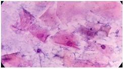
Fig. 1
Simply explained Pap Smears of benign pockets of retraction show only cuticular cell elements. However, as demonstrated by Fig.1 those pockets of retraction that had the tendency to become invasive and destructive, demonstrated an increased number of desquamated cells with protoplasms.
MICROCAUTHERIZATION
At the time of these studies, and in spite of having excellent means of visualization provided by the Surgical Microscope, there was not a tool available that could be used in the excision of these pockets of retraction, using minimally invasive approaches.
This fact prompted us to focus our attention to develop equipment that could be used to accomplish these goals.
After some experimentation, it became quite clear that the simplest and most economical way to resolve this problem was by using FOCAL HEAT.
Although heat and cauterization had been used since time immemorial, it never had been used in small confined areas such as the ear canal.
Furthermore, to our knowledge, nobody had ever built a miniature cautery that could be used in confined areas, such as the ear canal.
These problems were resolved by constructing a miniature cautery, with a milimetric tip, and with a built in heat sink, that will immediately draw the heat away upon deactivation.
In addition the microcomputer helped resolve many problems in the ear, nose, and throat field. Many of these publications, still available on the web, attest to this fact.
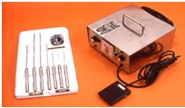
Fig 2. Kirchner Microcautery kit .Showing the Power Pack and Laryngeal (Adult and infant) and otological probes.
EXICION AND RECONSTRUCTION OF TYMPANIC MEMBRANE POCKETS OF COLLAPSE USING THE KIRCHNER MICROCAUTERY.
The following figures demonstrate how pocket of retraction are removed using the KIRCHNER MICROCAUTERY.
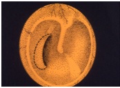 Fig. 3 Pocket of retraction. As seen under the otological Microscope |
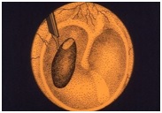 Fig. 4 Excision with Microcautery. |
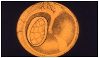 Fig. 5 Gel foam wedges holding the fascia Graft |
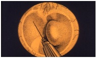 Fig. 6 |
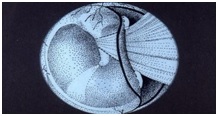 Fig. 7 |
Although the simplest type of pocket of retraction is shown here, there were other more complex instances where these pockets were found deeply retracted, behind the bony recess of the tympanic annulus. In these instances modified techniques were used, such as the eversion of this collapsed portion, by injection of saline solution in the middle ear. Fig 6 In these instances grafting was also modified as shown by slide Fig 7.
To end, one should mention, that all of these techniques, prevented any modification of the bony structure of the middle ear, particularly the annulus tympanicus. Because, without that structural support, the all important pneumatic space of the middle ear, is highly compromised.
Weekly Ads For pharmacy products.
Microcauterization in Otolaryngology
Fernando R. Kirchner, MD; Pedro S. Toledo, PhD
Arch Otolaryngol. 1974;99(3):198-202. doi:10.1001/archotol.1974.00780030206010.
 Intended for EAR HYGIENE: cleaning of debris, itch relief, exfoliation, water extraction, and superficial wax around the ear and outer ear canal. |
 NeilMed Clearcanal – Dr. Mehta’s ear wax removal complete kit |
(19177)
Rachel Momo liked this on Facebook.
Fernando Kirchner liked this on Facebook.
Julie Carrington liked this on Facebook.
Susan Stark Patti liked this on Facebook.
Julie Adams liked this on Facebook.
Where can I buy in Australia
Linda White Besink liked this on Facebook.
That is wonderful but where can I buy it in Ontario
Bhavna Garg liked this on Facebook.
Gilbert F. Jimenez liked this on Facebook.
Joanne Mercier-Moran liked this on Facebook.
Nevin Grushka liked this on Facebook.